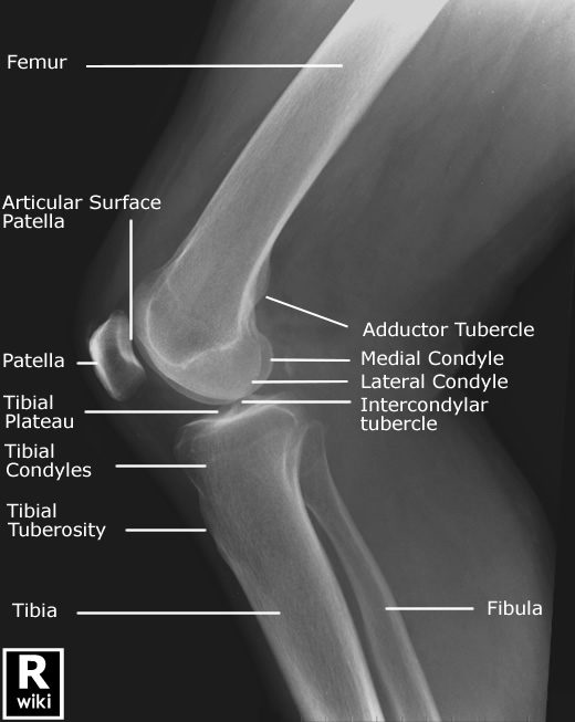
Left Knee Xray Labeled . Web the knee series is a set of radiographs taken to investigate knee joint pathology, often in the context of trauma. The picture shows the soft tissues and bones in and. Find out how to diagnose meniscal,. Learn about the bones, cartilage, ligaments, and common injuries of. This view demonstrates the distal femur and proximal tibia/fibula in their natural anatomical position allowing for assessment of.
from www.wikiradiography.net
Web the knee series is a set of radiographs taken to investigate knee joint pathology, often in the context of trauma. This view demonstrates the distal femur and proximal tibia/fibula in their natural anatomical position allowing for assessment of. Learn about the bones, cartilage, ligaments, and common injuries of. The picture shows the soft tissues and bones in and. Find out how to diagnose meniscal,.
Lateral Knee Radiography wikiRadiography
Left Knee Xray Labeled The picture shows the soft tissues and bones in and. Web the knee series is a set of radiographs taken to investigate knee joint pathology, often in the context of trauma. This view demonstrates the distal femur and proximal tibia/fibula in their natural anatomical position allowing for assessment of. Find out how to diagnose meniscal,. Learn about the bones, cartilage, ligaments, and common injuries of. The picture shows the soft tissues and bones in and.
From www.sexizpix.com
Knee X Ray Anatomy Procedure What To Expect Sexiz Pix Left Knee Xray Labeled Learn about the bones, cartilage, ligaments, and common injuries of. The picture shows the soft tissues and bones in and. This view demonstrates the distal femur and proximal tibia/fibula in their natural anatomical position allowing for assessment of. Find out how to diagnose meniscal,. Web the knee series is a set of radiographs taken to investigate knee joint pathology, often. Left Knee Xray Labeled.
From www.dreamstime.com
Xray of Human Knee Severe Osteoarthritis of the Knee Normal Ligaments Left Knee Xray Labeled Learn about the bones, cartilage, ligaments, and common injuries of. The picture shows the soft tissues and bones in and. This view demonstrates the distal femur and proximal tibia/fibula in their natural anatomical position allowing for assessment of. Find out how to diagnose meniscal,. Web the knee series is a set of radiographs taken to investigate knee joint pathology, often. Left Knee Xray Labeled.
From healthproadvice.com
Three Different Types of Knee XRays With Photos HealthProAdvice Left Knee Xray Labeled This view demonstrates the distal femur and proximal tibia/fibula in their natural anatomical position allowing for assessment of. The picture shows the soft tissues and bones in and. Learn about the bones, cartilage, ligaments, and common injuries of. Find out how to diagnose meniscal,. Web the knee series is a set of radiographs taken to investigate knee joint pathology, often. Left Knee Xray Labeled.
From mydiagram.online
[DIAGRAM] Diagram Of Normal Knee Left Knee Xray Labeled Find out how to diagnose meniscal,. This view demonstrates the distal femur and proximal tibia/fibula in their natural anatomical position allowing for assessment of. The picture shows the soft tissues and bones in and. Learn about the bones, cartilage, ligaments, and common injuries of. Web the knee series is a set of radiographs taken to investigate knee joint pathology, often. Left Knee Xray Labeled.
From www.pinterest.es
Xknee Startradiology Radiology student, Radiology, Medical anatomy Left Knee Xray Labeled Learn about the bones, cartilage, ligaments, and common injuries of. Find out how to diagnose meniscal,. Web the knee series is a set of radiographs taken to investigate knee joint pathology, often in the context of trauma. The picture shows the soft tissues and bones in and. This view demonstrates the distal femur and proximal tibia/fibula in their natural anatomical. Left Knee Xray Labeled.
From www.melbourneradiology.com.au
Bulk Billing Xrays Melbourne Melbourne Radiology Clinic Left Knee Xray Labeled Web the knee series is a set of radiographs taken to investigate knee joint pathology, often in the context of trauma. Find out how to diagnose meniscal,. Learn about the bones, cartilage, ligaments, and common injuries of. This view demonstrates the distal femur and proximal tibia/fibula in their natural anatomical position allowing for assessment of. The picture shows the soft. Left Knee Xray Labeled.
From radiopaedia.org
Image Left Knee Xray Labeled Learn about the bones, cartilage, ligaments, and common injuries of. This view demonstrates the distal femur and proximal tibia/fibula in their natural anatomical position allowing for assessment of. Find out how to diagnose meniscal,. The picture shows the soft tissues and bones in and. Web the knee series is a set of radiographs taken to investigate knee joint pathology, often. Left Knee Xray Labeled.
From www.ubicaciondepersonas.cdmx.gob.mx
Lateral Xray Of The Knee By Medical Body Scans ubicaciondepersonas Left Knee Xray Labeled Find out how to diagnose meniscal,. Learn about the bones, cartilage, ligaments, and common injuries of. Web the knee series is a set of radiographs taken to investigate knee joint pathology, often in the context of trauma. This view demonstrates the distal femur and proximal tibia/fibula in their natural anatomical position allowing for assessment of. The picture shows the soft. Left Knee Xray Labeled.
From www.cortho.org
Case Study Custom Left Knee Replacement in 66 yr. Old Male Left Knee Xray Labeled Find out how to diagnose meniscal,. Web the knee series is a set of radiographs taken to investigate knee joint pathology, often in the context of trauma. The picture shows the soft tissues and bones in and. Learn about the bones, cartilage, ligaments, and common injuries of. This view demonstrates the distal femur and proximal tibia/fibula in their natural anatomical. Left Knee Xray Labeled.
From www.cortho.org
Runners Knee New York Dr. Nakul Karkare Left Knee Xray Labeled Learn about the bones, cartilage, ligaments, and common injuries of. Find out how to diagnose meniscal,. Web the knee series is a set of radiographs taken to investigate knee joint pathology, often in the context of trauma. This view demonstrates the distal femur and proximal tibia/fibula in their natural anatomical position allowing for assessment of. The picture shows the soft. Left Knee Xray Labeled.
From www.orthobullets.com
Adult Knee Radiographic Views Trauma Orthobullets Left Knee Xray Labeled Learn about the bones, cartilage, ligaments, and common injuries of. Web the knee series is a set of radiographs taken to investigate knee joint pathology, often in the context of trauma. Find out how to diagnose meniscal,. This view demonstrates the distal femur and proximal tibia/fibula in their natural anatomical position allowing for assessment of. The picture shows the soft. Left Knee Xray Labeled.
From radiopaedia.org
Image Left Knee Xray Labeled This view demonstrates the distal femur and proximal tibia/fibula in their natural anatomical position allowing for assessment of. Find out how to diagnose meniscal,. The picture shows the soft tissues and bones in and. Web the knee series is a set of radiographs taken to investigate knee joint pathology, often in the context of trauma. Learn about the bones, cartilage,. Left Knee Xray Labeled.
From www.wikiradiography.net
Lateral Knee Radiography wikiRadiography Left Knee Xray Labeled This view demonstrates the distal femur and proximal tibia/fibula in their natural anatomical position allowing for assessment of. Web the knee series is a set of radiographs taken to investigate knee joint pathology, often in the context of trauma. The picture shows the soft tissues and bones in and. Find out how to diagnose meniscal,. Learn about the bones, cartilage,. Left Knee Xray Labeled.
From www.youtube.com
What Does a Knee X Ray Look Like? YouTube Left Knee Xray Labeled The picture shows the soft tissues and bones in and. Find out how to diagnose meniscal,. Learn about the bones, cartilage, ligaments, and common injuries of. Web the knee series is a set of radiographs taken to investigate knee joint pathology, often in the context of trauma. This view demonstrates the distal femur and proximal tibia/fibula in their natural anatomical. Left Knee Xray Labeled.
From exofttlev.blob.core.windows.net
What Does An X Ray Show Of The Knee at Anna Killinger blog Left Knee Xray Labeled Web the knee series is a set of radiographs taken to investigate knee joint pathology, often in the context of trauma. This view demonstrates the distal femur and proximal tibia/fibula in their natural anatomical position allowing for assessment of. The picture shows the soft tissues and bones in and. Find out how to diagnose meniscal,. Learn about the bones, cartilage,. Left Knee Xray Labeled.
From www.bmj.com
Lateral radiograph of the knee The BMJ Left Knee Xray Labeled Find out how to diagnose meniscal,. This view demonstrates the distal femur and proximal tibia/fibula in their natural anatomical position allowing for assessment of. Learn about the bones, cartilage, ligaments, and common injuries of. The picture shows the soft tissues and bones in and. Web the knee series is a set of radiographs taken to investigate knee joint pathology, often. Left Knee Xray Labeled.
From www.tamingthesru.com
Diagnostics Knee and Ankle Xrays — Taming the SRU Left Knee Xray Labeled This view demonstrates the distal femur and proximal tibia/fibula in their natural anatomical position allowing for assessment of. Find out how to diagnose meniscal,. The picture shows the soft tissues and bones in and. Learn about the bones, cartilage, ligaments, and common injuries of. Web the knee series is a set of radiographs taken to investigate knee joint pathology, often. Left Knee Xray Labeled.
From www.alamy.com
Normal Knee X Ray Stock Photos & Normal Knee X Ray Stock Images Alamy Left Knee Xray Labeled Learn about the bones, cartilage, ligaments, and common injuries of. Web the knee series is a set of radiographs taken to investigate knee joint pathology, often in the context of trauma. Find out how to diagnose meniscal,. The picture shows the soft tissues and bones in and. This view demonstrates the distal femur and proximal tibia/fibula in their natural anatomical. Left Knee Xray Labeled.
